




Description
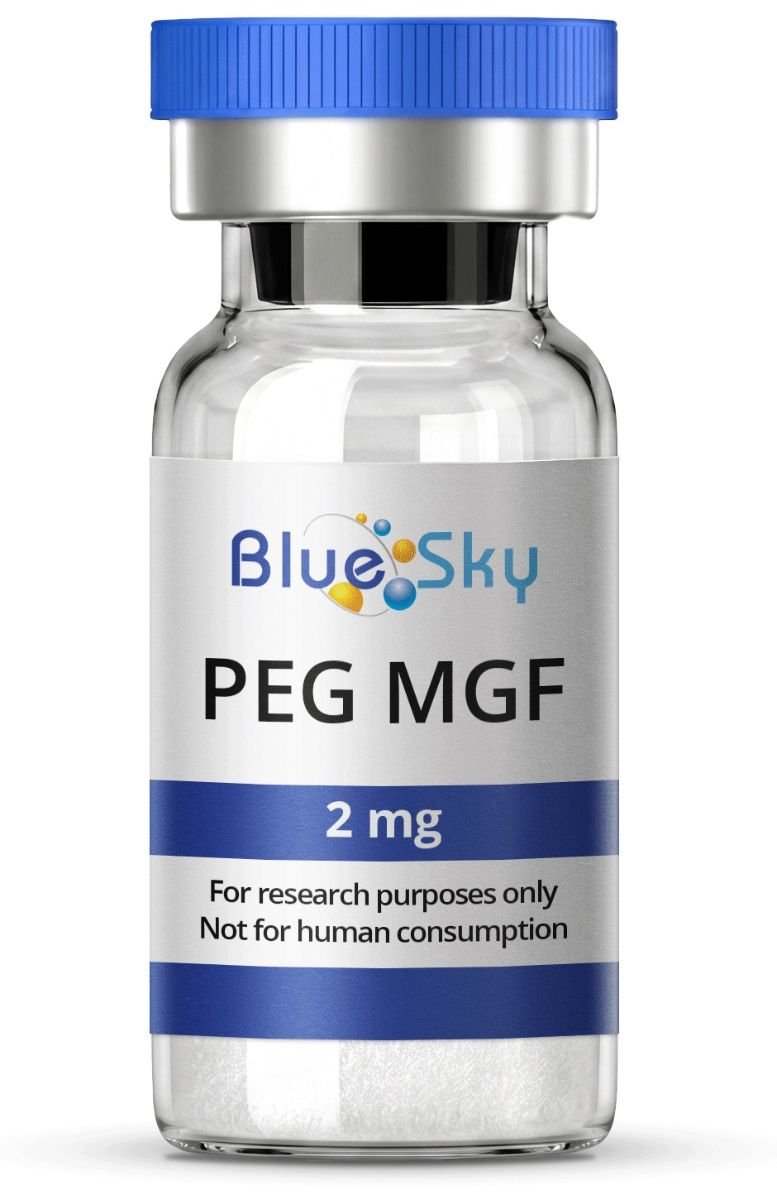
Buy PEG MGF 2 mg :
| Unit Size | 2 mg/vial |
| Unit Quantity | 1 vial |
| Purity (Mass Spectrometry and UV) | 99.64% |
| Sequence | PEG-Suc-YQPPSTNKNTKSQ(d)R(d)RKGSTFEEHKNH2 |
| Molecular Formula | C121H200N42O39 |
| Appearance | Lyophilized White Powder |
| Source | Chemical Synthesis |
| Storage | Lyophilized PEG MGF is stable at room Temperature for 90 days, however it is best to store in a freeze below – 8c for any extended period of time. |
| Terms | The products we offer are intended for laboratory research use only. Please familiarize yourself with our terms of service prior to ordering. |
PEG MGF 2mg
View Research Overview & References
PEG MGF is a synthetic analog of mechano-growth factor (MGF), a splice variant of insulin-like growth factor-1 (IGF-1). PEG MGF is created through a process called PEGylation, where polyethylene glycol (PEG) is attached to the molecule to extend its half-life by reducing rapid kidney clearance.1 This modification is studied for addressing MGF’s short half-life in laboratory research applications.
Muscle Cell Research
MGF is studied for its expression in skeletal muscle tissue and its role in stimulating myoblast growth and differentiation in models of injury or mechanical stress. In mouse model studies, direct injections of MGF into muscle tissue were associated with changes in recovery markers, oxidative stress markers, and inflammatory signaling.2,3
Cardiac Cell Research
Research into PEG MGF has also examined cardiac cell models. In vitro studies using microrods coated with MGF and PEG, designed to mimic cardiac tissue stiffness, examined mesenchymal stem cell migration toward MGF in this model.4 Separate studies examined MGF’s effects on cell death markers in neonatal rat ventricular cell models.
Cartilage Cell Research
Further studies have examined PEG MGF’s effects on chondrocyte migration in laboratory models, including research into MGF’s association with chondrocyte migration from bone to cartilage tissue.5
Important Notice
PEG MGF is intended exclusively for in vitro and in vivo (animal) laboratory research purposes and is not for human or animal consumption, diagnostic use, or therapeutic use. These findings do not establish safety, efficacy, or suitability for any human or animal application, and have not been evaluated by the FDA or any regulatory agency.
Summary
PEG MGF continues to be studied in vitro and in animal models for its effects on muscle, cardiac, and cartilage cell behavior. These findings are limited strictly to laboratory research settings.
References
1. Veronese FM, Mero A. The impact of PEGylation on biological therapies. BioDrugs. 2008;22(5):315-29.
2. Sun K, Cheung K, Au SWN, et al. Overexpression of Mechano-Growth Factor Modulates Inflammatory Cytokine Expression and Macrophage Resolution in Skeletal Muscle Injury. Front Physiol. 2019;9:999.
3. Liu X, Zeng Z, Chao L, et al. Impaired Skeletal Muscle Regeneration Induced by Macrophage Depletion Could Be Partly Ameliorated by MGF Injection. Front Physiol. 2019;10:601.
4. Doroudian G, Pinney J, Ayala P, et al. Sustained delivery of MGF peptide from microrods attracts stem cells and reduces apoptosis of myocytes.
5. Jing X, Ye Y, Bao Y, et al. Mechano-growth factor protects against mechanical overload-induced damage and promotes migration of growth plate chondrocytes through the RhoA/YAP pathway. Exp Cell Res. 2018;366(2):81-91.
Certificate of Analysis (COA) and third-party lab reports are available on request. Please contact our team for documentation specific to your lot/batch.